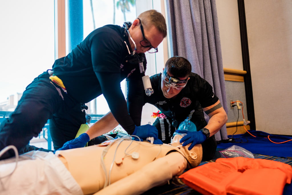
File Photo
An Overview of Three Uncommon Conditions
On the go? Listen to the article in the player below!
Introduction
Respiratory distress is one of the most common emergencies resulting in a 911 call. Whether it be an asthma attack, chronic obstructive pulmonary disease (COPD) exacerbation or complication of pneumonia, difficulty breathing is a symptom that prehospital providers encounter quite frequently. Most patients that call for emergency medical services for breathing problems have known-disease pathologies that are respiratory in nature. For example, patients with asthma, an obstructive pulmonary disorder, often present with chest tightness, wheezing and difficulty breathing. Nebulized albuterol and supplemental oxygen are normally first-line interventions for the typical asthma attack. Providers understand the mechanism of action for those medications since they aim to correct the impaired respiratory processes as a result of disease.
However, some patients that call 911 for difficulty breathing may not have conditions such as asthma, COPD or pneumonia. Certain disease processes that are neurological or muscular in nature may cause a patient to experience respiratory distress. Although rare, conditions such as esophageal achalasia, Guillain-Barré syndrome and amyotrophic lateral sclerosis (ALS) are non-respiratory processes that can cause a patient to have airway compromise. Prehospital providers should be aware of these types of conditions that result in breathing problems since the course of treatment provided may be atypical and airway management may be more difficult.
Esophageal Achalasia
Esophageal achalasia is the first rare condition that will be explored. Esophageal achalasia is also known as achalasia, cardiospasm, dyssynergia esophagus, esophageal aperistalsis and megaesophagus.1 It is a rare disorder of the esophagus and is characterized by impaired peristalsis, or the involuntary contraction and relaxation of esophageal muscles that creates wave-like movements to progress food down the esophagus. The ring-shaped muscle at the bottom of the esophagus is called the lower esophageal sphincter. Under normal conditions, it contracts and relaxes to move food through the esophagus. In achalasia, it fails to relax, causing a build-up of food in the esophagus and subsequent dilation.
Most patients with the disease typically present with dysphagia, or difficulty swallowing, as well as weight loss and regurgitation. Patients with achalasia are also more prone to aspirating saliva and food, which may cause pneumonia. Achalasia can be diagnosed by several methods. X-ray examination, especially with the use of barium, may indicate dilation of the esophagus and any retention of food or secretions. Other tests include esophageal manometry, which measures peristalsis, and upper endoscopy.2
Related: Optimal Prehospital Airway Management Depends on EMS System Efficiency
How does this disorder of the esophagus cause respiratory distress? When the esophagus massively dilates due to the back-up of un-swallowed food, it can cause tracheal compression, which occludes the airway. Symptoms of central airway compression typically arise after eating. Symptoms may include neck swelling and drooling caused by anterior displacement of the larynx.3 Patients may present with stridor or wheezing if the esophageal dilation is compressing the upper or lower airway. These respiratory complications of megaesophagus are often initially misdiagnosed as asthma.
However, typical interventions for asthma exacerbations are essentially ineffective in respiratory distress due to achalasia. Insertion of a nasogastric tube to minimize the volume of retained food material in the esophagus may reduce airway compression and thus mitigate respiratory distress. Other temporary interventions include the inhalation of a helium-oxygen gas mixture known as heliox as well as the administration of sublingual nitroglycerin to attempt to reduce lower esophageal sphincter tension.4 Racemic epinephrine may also be administered and has successfully reduced respiratory distress in some cases, especially in the prehospital environment.
Airway compromise is a rare complication of achalasia. Although rare, respiratory distress still occurs in some cases and can have dire consequences for a patient suffering from the disease. In the context of the prehospital environment, esophageal achalasia is not a common field diagnosis that most providers may propose unless the patient is able to specifically provide that information. However, providers should not take the “respiratory distress” dispatch at face value and instead should consider other causes of airway compromise that do not necessarily originate in the airway.
Guillain-Barré Syndrome
An additional non-respiratory disease pathology that may cause airway compromise is Guillain-Barré (GB) syndrome. GB is a rare neurological disorder that is characterized by one’s own immune system attacking the peripheral nervous system, or the network of nerves located outside of the brain and spinal cord. The affected patient’s immune system begins to associate its own nerve cells with an invading virus or bacteria and mounts a response, causing destruction of healthy cells.
Specifically, the myelin sheath, or the insulating layer that surrounds nerve cells and serves to protect and allow for faster propagation of electrical impulses, is damaged. The axon, or portion of the nerve cell that carries nerve impulses away from the cell body, may also be damaged. Destruction of either nerve cell component results in poor electrical signal transmission from nerve cell to nerve cell, which subsequently causes the muscles to lose their ability to respond to commands from the brain.5
Initial clinical manifestations of GB syndrome include unexplained sensations, such as tingling in the hands and feet, as well as pain in the legs or back.5 These symptoms are usually temporary and subside prior to more severe effects that are also longer-lasting. Global muscle weakness develops and may first present as difficulty performing ordinary tasks, such as walking or climbing stairs. Symptoms such as impaired vision, coordination issues and unsteadiness, difficulty swallowing, speaking or chewing may also arise. Abnormalities in cardiac rate and rhythm as well as blood pressure are possible complications as well. An important characteristic of GB syndrome is that symptoms occur bilaterally, meaning that both sides of the body exhibit deficits.
As days or weeks progress, most patients with the disease experience progressive weakness that can ultimately lead to partial or complete paralysis. In these instances, the muscles involved in inspiration and expiration (breathing) are affected, ultimately interfering with the body’s perfusion status. Patients who experience complete collapse of respiratory reflexes require aggressive airway management, including intubation and ventilation.
Related: A Modern Approach to Basic Airway Management
Guillain-Barré syndrome is usually diagnosed in its later stages, as the initial symptoms of the disease are non-discrete and may mimic various other pathologies. Physicians use a variety of physical and laboratory examination techniques in addition to obtaining a thorough patient history and timeline of symptom onset and progression to diagnose GB syndrome. Deep tendon reflexes such as the knee-jerk reaction are tested, with absent or diminished results used as the key diagnostic finding. Nerve conduction velocity tests are performed to measure the speed at which electrical nerve impulses travel by stimulating the nerve with electrode patches attached to the skin. Additionally, a spinal tap or lumbar puncture may be performed to detect any changes in the composition of the cerebrospinal fluid. Researchers have found that the CSF of patients with Guillain-Barré syndrome contains more protein than usual as well as a diminished white blood cell count.
Treatment of Guillain-Barré syndrome is multi-faceted. Acute interventions may be employed to interrupt immune-related nerve damage. One such intervention is plasma exchange, or plasmapheresis, which involves removing the patient’s blood from the venous circulation and extracting the plasma before returning it back to the patient. Blood plasma contains antibodies, some of which may be contributing to the autoimmune response, so plasmapheresis acts to remove the nerve-damaging antibodies and reduce the severity of the disease.
Another acute therapy used to treat Guillain-Barré syndrome is high-dose immunoglobulin therapy, or IVIg. Immunoglobulins are special proteins produced by the immune system to aid in infection mitigation. IVIg therapy provides these proteins to the patient in the form of an IV injection. IVIg therapy can both reduce the severity and duration of the disease and lower the level of effectiveness of harmful antibodies that attack the nerves. Supportive care in the form of monitoring perfusion status and airway management is also employed in Guillain-Barré patients. Upon improvement, many patients are transferred to external facilities for rehabilitation services and occupational therapy, as GB can affect a patient’s ability to perform mundane bodily functions such as walking or swallowing in the long term.6
Like esophageal achalasia, the exact cause of Guillain-Barré syndrome is unknown. However, it is known that the disease is not contagious or heritable in nature. One proposed theory to explain the underlying cause of GB syndrome is referred to as the “molecular mimicry/innocent bystander” theory. This theory states that molecules on some nerve cells share similarities with mimic molecules on some foreign microorganisms. When those microbes cause infection, the components of the immune system correctly respond. However, the immune system mistakenly attacks the myelin as well since it is similar in appearance to the infective agent. In other words, the immune system does not differentiate between self and non-self. This theory may be substantiated by the idea that Guillain-Barré syndrome is preceded by a viral or bacterial infection, which may result in a change in nerve cell composition. Additionally, the preceding infective event may make the immune system less discriminating and prevent it from recognizing its own nerves. Despite this theory, research is still ongoing regarding the specific etiology of Guillain-Barré syndrome.6
In the prehospital environment, Guillain-Barré syndrome is usually not a common differential diagnosis. However, paying attention to a patient’s history, including a recent viral or bacterial illness and noting the progression of symptoms, may assist in diagnosing the autoimmune disease, especially if the patient has no intact airway reflexes. As mentioned previously, the airway may be compromised by disease processes that are typically not associated with the respiratory system, which providers in the prehospital environment should be mindful of.
Amyotrophic Lateral Sclerosis
A third non-respiratory disease pathology that has the potential to complicate airway management is amyotrophic lateral sclerosis (ALS). ALS is a progressive neurodegenerative condition that is characterized by the gradual breakdown of nerve cells that control voluntary muscle movement. The actions of voluntary muscles consist of chewing, walking and talking among others. ALS is defined by the deterioration and ultimate death of motor neurons, which are nerve cells that extend from the brain to muscles throughout the body. Two types of motor neurons affected by ALS include the upper motor neurons and lower motor neurons. The upper motor neurons are located in the brain and transmit messages to motor neurons in the spinal cord and motor nuclei of the brain, referred to as lower motor neurons, and from the spinal cord and motor nuclei of the brain to a specific muscle or muscle group. The motor neurons are necessary for impulses to be transferred from the brain to muscles, causing voluntary movement. Therefore, the degeneration of upper and lower motor neurons affects the transmission of nerve impulses to the muscles. The absence of stimulation causes the muscles to gradually weaken, fasciculate or twitch, and atrophy or waste away. Ultimately, the brain loses complete control over voluntary movement.7
Early symptoms of ALS are typically muscle weakness or stiffness and may affect a variety of muscle groups, such as those responsible for grasping objects or chewing and swallowing. The muscle weakness is often gradual, painless and progressive. Other early signs of the disease vary but can consist of difficulty balancing, slurred speech or uncontrollable bouts of laughter or crying. Interestingly, patients with ALS have intact sensory neurons, so their senses of sight, touch, hearing and taste are not affected. Like Guillain-Barré syndrome, the muscles involved in breathing become affected in the end-stages of ALS, requiring permanent ventilatory support.
There are several potential risk factors for ALS, despite it affecting individuals of all races and ethnicities. The disease can strike at any age but most commonly occurs between the ages of 55-75. Men are more likely to develop ALS than women, yet this statistic essentially disappears with increasing age. Individuals of Caucasian and non-Hispanic descent are more likely to develop ALS. The majority of cases of ALS are sporadic, or at random, with no clear associated risk factors or familial history of the disease. The remaining percentage of cases are heritable in nature, with one parent required to carry the gene responsible for ALS.
Like Guillain-Barré syndrome, ALS is often difficult to diagnose. Physicians primarily rely on the patient’s history, physical and neurological exams, as well as a series of tests that rule out mimic disorders. Physical and neurological exams are also performed at regular intervals to detect any deteriorations or progressive symptoms that characterize the disease. A special imaging test called electromyography (EMG) detects electrical activity within muscle fibers and can aid in diagnosing ALS. Nerve conduction studies may also be performed to assess the ability of the patient’s nerves to transmit electrical impulses and evoke a response from the appropriate muscle. MRIs and bloodwork may also be performed in order to rule out other disorders.
There is no cure for ALS. Current treatments are supportive in nature and aim to manage symptoms and prevent unnecessary complications of the disease. Treatment may consist of services from physicians, pharmacists; physical, occupational and speech therapists – as well as nutritionists, social workers, psychologists and hospice nurses.8 Additionally, breathing support is also crucial to the treatment plan for ALS patients. Several resources are provided to ALS patients to improve breathing quality and other airway reflexes that may be affected by muscle weakness, such as the cough reflex.
Non-invasive ventilation (NIV) is a treatment that delivers breathing support in the form of a mask that the patient wears over the nose and/or mouth. NIV may be used only at night but may be required full-time as the patient’s perfusion status diminishes. Several techniques are also provided to patients to increase their ability to cough. Breath stacking is a technique in which an individual takes a series of small breaths without exhaling until the lungs are full before briefly holding the breath and then expelling the air with a cough. This allows for the ALS patient to maintain a cough reflex despite muscle weakness. When the muscles that control breathing are completely weakened, the patient may require mechanical ventilation via endotracheal intubation or tracheostomy.
Caring for ALS patients in the prehospital environment may be rare, but they still may call for emergency services due to difficulty breathing as a result of their progressive condition. Not only can ALS patients present with respiratory distress, but they may also require special management of pre-existing airway devices. In other words, they may require deep tracheal suctioning through their tracheostomy tube, or adjusted ventilator settings to promote adequate ventilation and perfusion. Regardless, providers should keep in mind that ALS patients encountered in the field require special management and considerations due to the progressive nature of the condition and its critical effect on the airway.
Conclusion
Despite the commonality of a “difficulty breathing” 911 call, asthma, COPD, or an allergic reaction are not the only causes of the symptom that creates distress and prompts a patient or family member to request EMS. There are a multitude of disease processes, although uncommon, that are not respiratory in nature that can complicate airway management. Esophageal achalasia, Guillain-Barré syndrome, and ALS are just three examples of conditions in which respiratory failure is possible despite having muscular, autoimmune and neurological origins. The prehospital provider should be mindful of such diseases as they require special considerations and in some cases completely different treatment plans than the typical respiratory distress call. The maintenance of a patent airway is essential to patient care and there is a wide scope of etiologies beyond common respiratory conditions that threaten it.
References
1. “Achalasia.” NORD (National Organization for Rare Disorders), 14 Aug. 2020a, rarediseases.org/rare-diseases/achalasia/.
2. Spechler , Stephen J. “Patient Education: Achalasia (Beyond the Basics).” UpToDate, 22 May 2019, www.uptodate.com/contents/achalasia-beyond-the-basics/print.
3. Adamson, Rosemary, et al. “Acute Respiratory Failure Secondary to Achalasia .” Annals of the American Thoracic Society, June 2013, pp. 268–271.
4. “Achalasia.” Mayo Clinic, Mayo Foundation for Medical Education and Research, 21 Oct. 2020b, www.mayoclinic.org/diseases-conditions/achalasia/diagnosis-treatment/drc-20352851.
5. “Guillain-Barre Syndrome.” Mayo Clinic, Mayo Foundation for Medical Education and Research, 17 Sept. 2020, www.mayoclinic.org/diseases-conditions/guillain-barre-syndrome/symptoms-causes/syc-20362793.
6. “Guillain-Barré Syndrome Fact Sheet.” National Institute of Neurological Disorders and Stroke, U.S. Department of Health and Human Services, 16 Mar. 2020, www.ninds.nih.gov/Disorders/Patient-Caregiver-Education/Fact-Sheets/Guillain-Barr%C3%A9-Syndrome-Fact-Sheet#3139_1.
7. “What Is ALS?” The ALS Association, www.als.org/understanding-als/what-is-als.
8. “Amyotrophic Lateral Sclerosis (ALS) Fact Sheet.” National Institute of Neurological Disorders and Stroke, U.S. Department of Health and Human Services, June 2013, www.ninds.nih.gov/Disorders/Patient-Caregiver-Education/Fact-Sheets/Amyotrophic-Lateral-Sclerosis-ALS-Fact-Sheet.
Kelly Ward has a bachelor’s degree in emergency medicine from the University of Pittsburgh and is a practicing paramedic. She is currently in nursing school and hopes to combine her interests in critical care and prehospital emergency medicine by pursuing a career in flight nursing.


Recent Comments