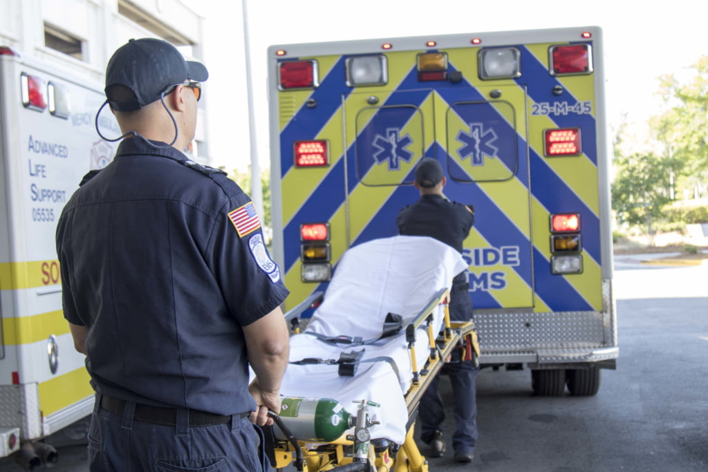
Paramedics with Southside EMS prep the ambulance after transporting a patient to Memorial Hospital in Savannah, Georgia May 3, 2017. (US Army photo by Staff Sgt. Kellen Stuart, 3rd CAB Public Affairs)
In previous columns, we’ve reviewed various non-invasive ventilation (NIV) modalities: CPAP, BiPAP and High-Flow Nasal Cannula. While there are similarities, each has a unique niche in management of respiratory distress. This column will compare and contrast NIV modalities and highlight advantages, disadvantages and selection criteria that will help you make the best choice for improving patient outcomes. Let’s review three case studies.
Case #1
You are called to see a 78-year-old male who has been experiencing increasing shortness of breath over the past 24 hours. He has a history of heart failure (with an ejection fraction of 30%) as well as atrial fibrillation. His medications include carvedilol, captopril, spironolactone, dabigatran and home oxygen at 2 LPM which he increased to 4 LPM prior to calling EMS. Vitals signs show an irregular pulse of 118, respirations of 32 and labored, blood pressure 164/88 and oxygen saturation of 78% on 4 liters by nasal cannula. He has peripheral cyanosis, crackles two-thirds of the way up from both bases, and is profusely diaphoretic, but afebrile. The 12-lead ECG shows uncontrolled atrial fibrillation with no evidence of acute ischemia. The EtCO2 is 62 mmHg.
Given the history, this is likely acute pulmonary edema (APE) which responds nearly instantaneously to CPAP. In fact, the earlier CPAP is applied in APE, the better the outcomes. With the application of CPAP at 10 CWP by BLS first responders, the patient improved within minutes. On arrival of the ALS transport unit, his heart rate was 94, respirations 28, blood pressure 138/70, oxygen saturation 96% and EtCO2 50. ALS continued the CPAP, provided IV bolus nitroglycerin and a nitro infusion during transport. After three days in a telemetry unit, the patient was discharged home. Conversely, had the BLS responders applied high-flow oxygen (usually with a non-rebreather mask), the patient would likely have continued to deteriorate, developing pink frothy sputum, decreased level of consciousness and progressed to respiratory arrest, requiring bag-valve-mask ventilation and endotracheal intubation by the ALS transport unit. Mechanical ventilation and sedation require ICU care, often complicated by ventilator associated pneumonia (VAP). Deconditioning and prolonged hospitalization often lead to other complications.
While APE often presents with impressively low oxygen saturations, oxygenation is not the primary problem, but rather acute decompensated heart failure (ADHF). In APE, one or both ventricles cannot overcome the resistance they are ejecting against. The reduction in transpulmonary pressures seen following application of CPAP powerfully lowers afterload, leading most APE patients to improve within seconds.1-2 The combined effects of CPAP improving oxygenation from splinting open terminal airways adds to clinical improvement.
Case #2
You are called to see a 64-year-old male with increasing shortness of breath over the past three days. He has a history of COPD. Home medications include albuterol PRN, prednisone, Combivent® and home oxygen at 3 LPM. He has no allergies. Vital signs show a regular heart rate of 138, respirations of 38 and labored, blood pressure of 148/80 and oxygen saturation of 93% on 3 LPM nasal cannula. The patient is febrile (101.5 °F), has purulent sputum, scattered rhonchi across all lung fields, and is using tripod posturing to breathe. There is no edema. The 12 lead shows sinus tachycardia with no evidence of acute ischemia. His EtCO2 is 88.
This gentleman is not acutely hypoxic but is significantly hypercarbic and, judging from the clinical picture, very close to, if not already in, respiratory failure. His tachypnea and high EtCO2 suggest that he needs assistance breathing. While CPAP alone may help assist his tired respiratory musculature, the pressure applied continuously throughout the respiratory cycle requires increased effort to exhale and may be intolerable. BiPAP delivers two different pressures, an inspiratory positive airway pressure (IPAP) and a lower, expiratory positive airway pressure (EPAP). The lower pressure during expiration reduces the work of exhaling. The difference between IPAP and EPAP is called Pressure Support (PS) or Delta P, and directly correlates to tidal volume. The greater the difference in pressures, the higher the patient’s tidal volume will be and, as a result, CO2 levels decrease. The ability of BiPAP to increase tidal volumes, therefore lowering CO2 levels, is what differentiates it from CPAP.3-4
The history and clinical picture strongly suggests pneumonia leading to an exacerbation of the patient’s COPD. To support breathing, CPAP was immediately initiated at 10 CWP using a disposable, single use unit with an in-line nebulizer of albuterol and ipratropium. Once in the ambulance, the patient was more comfortable but his oxygen saturation had dropped to 89% and his EtCO2 had climbed to 95. A transport ventilator circuit was connected to the CPAP mask and BiPAP was initiated with an IPAP of 10, an EPAP of 4 CWP and an FiO2 of 0.4. The patient became markedly more comfortable, and reported a significant decrease in dyspnea. After 15 minutes, his heart rate was 98 and regular, respiratory rate 28, BP 130/72, SpO2 96% and EtCO2 77.
For patients who are tiring from increased work of breathing due to airflow limitations from causes such as pneumonia, chest wall injuries, asthma, ALS, etc., BiPAP not only supports respirations but eases the work of expiration, helping to more efficiently clear CO2.
Case #3
You are called by family to see a 64-year-old female with terminal liver cancer that has multiple metastases throughout her body. The family believes the patient is having difficulty breathing. She has a valid do not intubate/do not resuscitate order. Medicine include morphine PRN, lorazepam PRN, home oxygen at 2 LPM PRN. She is allergic to sulfa antibiotics and amlodipine. Vitals signs include a heart rate of 115 and regular, respirations are 28 and regular, blood pressure of 100/50, and SpO2 80% on 2 liters of oxygen by nasal cannula. The patient is restless but otherwise somnolent, responding to loud voices and tapping on the shoulder. She is cachexic, and seems to be dyspneic, but is not able to answer questions coherently. The family tells you that her mental state is at baseline. Her EtCO2 is 45; a 12 lead reveals sinus tachycardia, with no evidence of ischemia. The rest of your exam is unremarkable.
High-flow nasal cannula (HFNC) is ideal for hypoxemic respiratory failure5-6, as is the case with this terminally ill patient. Increasingly used in hospital settings, HFNC uses flow rates of up to 60 LPM and FiO2’s of up to 100%. The gas is heated and humidified for comfort and delivered through a large bore, soft nasal cannula. While elevated CO2 is the usual cause of air hunger, significant hypoxia can also cause dyspnea, especially when saturations dip into low 80s and hypoxia can certainly lead to restlessness. Since this patient’s CO2 is normal and breath sounds clear, you should attempt to improve her oxygenation and evaluate whether that helps reduce her restlessness and apparent dyspnea.
HFNC equipment is not yet available in the outpatient setting but high FiO2s can be achieved with a combination of nasal cannula and non-rebreather mask. Prolonged nasal cannula flow rates above 6 LPM can be irritating and their drying effect on the mucus membranes may lead to nosebleeds. Home oxygen can be humidified to reduce this risk. CPAP using a disposable device is unlikely to help this patient as the inspired FiO2 is not likely to be as high as a non-rebreather mask would provide owing to the venturi design used to achieve the desired CPAP pressures. After adjusting the oxygen flow, you are able to achieve an SpO2 of 92% with 6 LPM of oxygen by nasal cannula. Repeat vital signs show a heart rate of 88, respirations of 21 and regular, blood pressure 98/46, SpO2 92% and EtCO2 44. The home oxygen unit is humidified and the patient now appears comfortable. You contact the hospice nurse on call and advise him of your findings and treatment.
Aside from CPAP, there are few clinical guidelines or protocols addressing the indications and use of NIV. Use of NIV is both an art and a science. Familiarity with the etiologies of respiratory distress and relative advantages and disadvantages of each type of NIV will help you to choose a device most appropriate for your patient.
References
- Weng CL, Zhao YT, Liu QH, et al. Meta-analysis: noninvasive ventilation in acute cardiogenic pulmonary edema. Ann Intern Med. 2010;152:590-600.
- Dhainaut JF, Devaux JY, Monsallier JF, et al. Mechanisms of decreased left ventricular preload during continuous positive pressure ventilation in ARDS. Chest. 1986;90:74-80.
- Ferguson GT, Gilmartin M. CO2 rebreathing during BiPAP ventilatory assistance. Am J Respir Crit Care Med 1995; 151:1126.
- Liesching T, Kwok H, Hill NS. Acute applications of noninvasive positive pressure ventilation. Chest 2003; 124:699.
- Mauri T, Alban L, Turrini C, et al. Optimum support by high-flow nasal cannula in acute hypoxemic respiratory failure: effects of increasing flow rates. Intensive Care Med 2017; 43:1453.
- Rochwerg B, Granton D, Wang DX, et al. High flow nasal cannula compared with conventional oxygen therapy for acute hypoxemic respiratory failure: a systematic review and meta-analysis. Intensive Care Med 2019; 45:563.
Mike McEvoy, PhD, NRP, RN, CCRN, is the EMS coordinator for Saratoga County, New York, and the professional development coordinator for Clifton Park and Halfmoon Ambulance. He is a nurse clinician in the adult and pediatric cardiac surgery intensive care units at Albany Medical Center, where he also teaches critical care medicine. McEvoy is the chief medical officer and firefighter/paramedic for West Crescent Fire Department in Clifton, New York. He is also the chair of the EMS Section board of directors for the International Association of Fire Chiefs and a member of the New York State Governor’s EMS Advisory Council. He is a lead author of the textbook Critical Care Transport, the “Informed” Pocket References (Jones & Bartlett), and the American Academy of Pediatrics textbook Pediatric Education for Prehospital Professionals (PEPP).


Recent Comments